Generating Hope
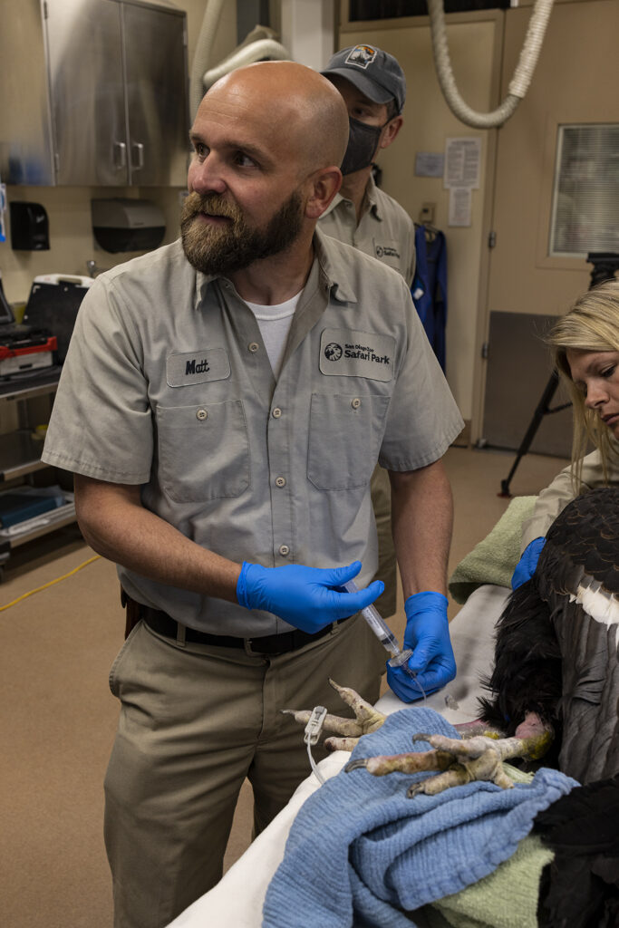
BY Matthew E. Kinney, DVM, DACZM
Senior Veterinarian for San Diego Zoo Wildlife Alliance
Regenerative Medicine
in Veterinary Care
Regenerative medicine is a relatively new approach to patient care that is focused on the repair, replacement, or regeneration of cells, tissues, or organs to restore impaired function, from a variety of causes. The veterinary staff at San Diego Zoo Wildlife Alliance uses regenerative medicine techniques in a variety of clinical cases, with a focus on one particular area of regenerative medicine: mesenchymal stem cells.
Going Cellular
Mesenchymal stem cells have the capacity for self-renewal, and can differentiate into a number of different cells. These types of stem cells can be collected from fat and umbilical cord tissue, shortly after the birth of an animal.
Our wildlife health teams have not only embraced regenerative medicine, but have become leaders in the use of this non-traditional modality. Over two decades ago, mesenchymal stem cells were first used at the San Diego Zoo Safari Park following fat collection, cell isolation, and re-administration into a zebra that was diagnosed with orthopedic disease, causing hind limb limping.
The improvements observed in that case resulted in veterinarians further exploring the treatment possibilities offered, by translating techniques and indications for use from domestic animal veterinary medicine and human medicine. Since initial use over two decades ago, our wildlife health teams have been both preparing for potential cases and utilizing mesenchymal stem cells in a variety of clinical cases.
An Early Start
The plan to use mesenchymal stem cells is often initiated well in advance of their intended use—in some instances, even before the patient is born! Cells contained within the umbilical cord have a high percentage of mesenchymal stem cells, and therefore this tissue is a great source to collect from for further storage and clinical use. In many instances, wildlife health and care teams will work together closely after an animal is determined to be pregnant, to plan for the collection of the umbilical cord—which is naturally separated from the offspring shortly after birth. The actual collection and processing of the cord can be a challenge, as the tissue must be collected safely and without disrupting the bond between the offspring and mother. Once collected, the cord is cleaned, and specific sections are taken for cell harvesting at a laboratory in the San Diego area.
After a few weeks, the cells are cultured, expanded, and frozen for clinical use. These cells can be stored for years until a clinical use arises, at which time they are unfrozen and can be administered to a patient within a day or two, which is often faster than some medicines can be ordered and delivered. Mesenchymal stem cells are not specific to individuals, so one individual within a species may be the “donor,” but their cells can be administered to herd or flock mates, if needed.
Good Fat
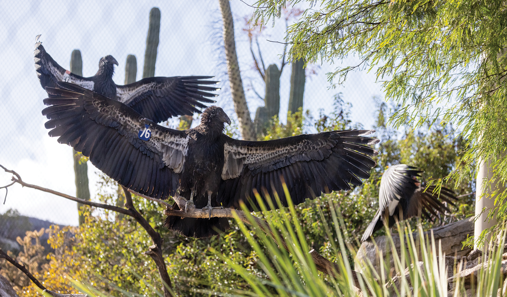
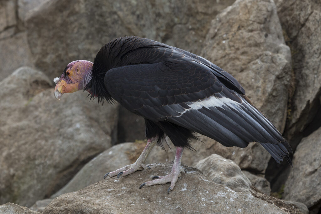
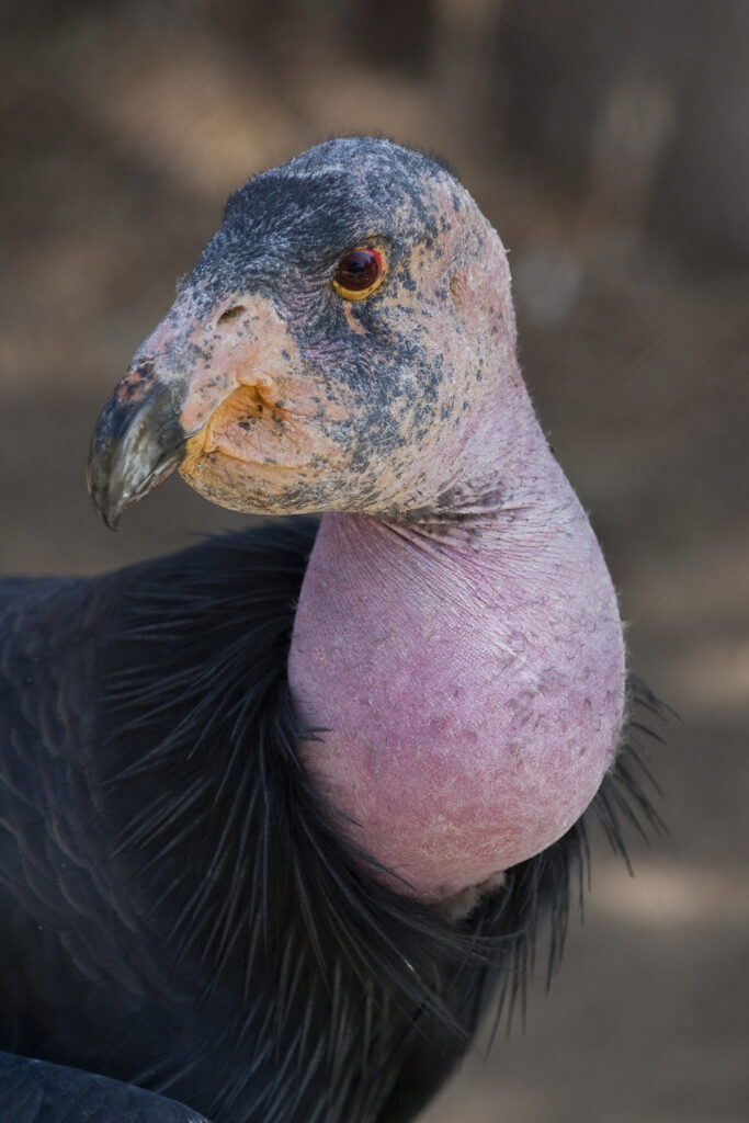
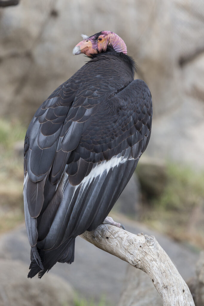
In addition to obtaining cells from umbilical tissue, another common source is adipose tissue, or fat. Adipose tissue collection for cell harvesting does not require the birth of an offspring, and it can be collected from an animal of any age. Cheetah cells proved to be among the more difficult to collect, as cheetahs have minimal subcutaneous (underneath-the-skin) fat. However, a large amount of fat can be collected from the abdominal cavity, and this was obtained opportunistically in a cheetah at the Safari Park during a laparoscopic surgical procedure. In most other mammal species, fat can be found immediately beneath the skin, allowing easy sampling. In birds, fat can be a little more difficult to obtain, as the location of fat stores can vary greatly between species. The wildlife health team recently performed a computed tomography (CT) scan on a California condor, as part of a diagnostic evaluation, and to locate an area of abundant and accessible fat stores to collect for mesenchymal stem cell culture.
Just the Beginning
Once umbilical or adipose tissue is collected and mesenchymal stem cells are isolated, they can be used for clinical cases with very little advance notice. By far the most common use of regenerative therapy is for diseases affecting bones and cartilage that surround joints. In addition to regenerative capacity, cell-related factors likely influence the microenvironment of the diseased tissue, which may have direct anti-inflammatory and pain-relieving effects.
Mesenchymal stem cells have been used in a traditional application for degenerative joint disease on a variety of species at the San Diego Zoo and Safari Park, including giraffes, rhinos, and California condors. This modality has also been applied as a novel therapy for treatment of non-orthopedic-related disease in a variety of animals. Repeated intravenous administration of mesenchymal stem cells has been used in cheetahs with severe liver disease, and in African elephants with viral infections.
The use of regenerative medicine in zoological veterinary practice remains in its infancy. Thoughtful planning to maximize collection opportunities, identification of disease processes that may benefit from cell or tissue repair and regeneration using these cell types, and preparing cells well in advance of their anticipated use has allowed us to apply these modalities for timely and effective treatment in a variety of patients.