Views You Can Use
New CT Scanners Take Animal Care to the Next Level
BY Karyl Carmignani
Photography by Ken Bohn
Few things excite veterinarians, technicians, and keepers more than a non-invasive, highly precise machine that vastly improves animal health and welfare. Called computed tomography in medical parlance, it is also known as a CT scan (or CAT scan) to the rest of us. A large, doughnut-shaped machine takes scores of incredibly detailed, cross-sectional X-ray images (radiographs) at different angles of the body, which provide mind-blowing views of the inner workings of an animal (or person) and help guide effective medical treatment. A CT scan reveals intricate details of bones, organs, blood vessels, and soft tissue that cannot be seen in conventional X-rays. Veterinarians and other medical professionals use specialized equipment, software, and expertise to diagnose cancer, trauma, tumors, musculoskeletal disorders, and other abnormalities, with detail that can be life saving.
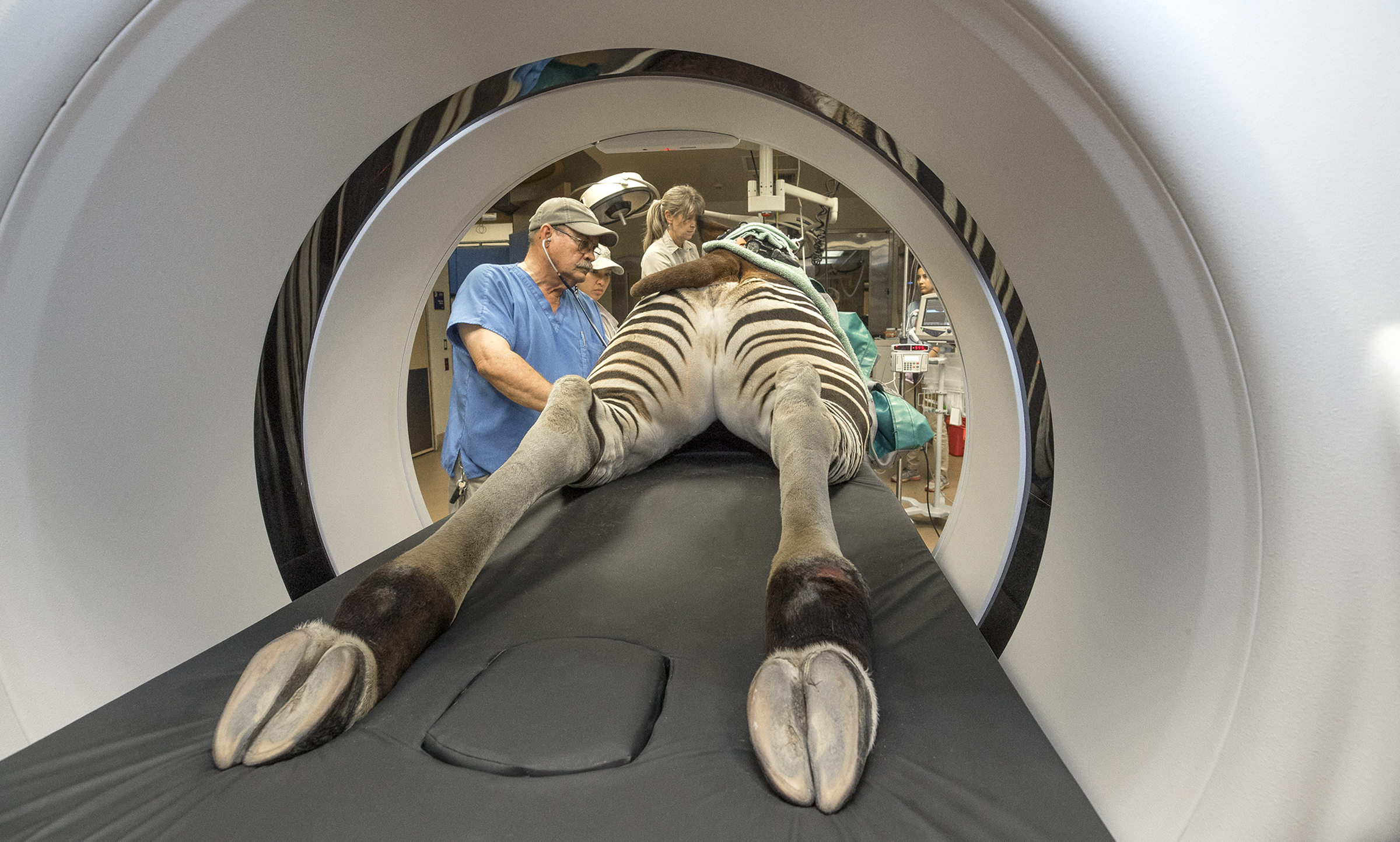
INSIDE LOOK
This young tiger under anesthesia is getting top-notch health care at the Safari Park.
What a “Bore”
Years ago, Pat Morris, DVM, San Diego Zoo, had the vision for the use of a CT scanner and understood how it would transcend our veterinary care. “Dr. Morris did a lot of the legwork and research about computed tomography long before we had the funds to get a scanner,” said Matt Kinney, DVM, San Diego Zoo Safari Park. “He was ahead of his time.” And Dr. Morris knew what we needed in a CT scanner: it had to be portable, have a large bore hole (gantry), and use sturdy, weight-bearing tables. Thanks to a generous donation in 2017 by the Alt Estate, the Safari Park was gifted the money to purchase a BodyTom Elite CT scanner by NeuroLogica, a subsidiary of Samsung. The Zoo also received a CT scanner, and both facilities get support and training from the helpful experts at Samsung, who are keen to see their product used in an animal-centric way.
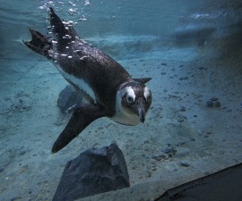
REVEALING
The CT scanner is a noninvasive way to reveal different views of the innermost workings of animals, like this otter.
“This scanner is unique in that it moves to the patient,” Dr. Kinney explained. In contrast, humans getting a CT scan are on a table and moved into the stationary machine. Once the animal is secured on the table (usually anesthetized), red lasers on the scanner aid staff in positioning the patient and the body area to be scanned. The 33-inch-wide (85-centimeter) opening (which can accommodate most animals in the collection) glides over the patient as the X-ray tube spins rapidly around the patient’s body. The machine sends out cone-shaped X-ray beams, and the rays strike a plate that “reads” the information—varying absorption rates of the varying thicknesses of tissue and bone—and transmits that data into a viewable format to a wireless workstation.
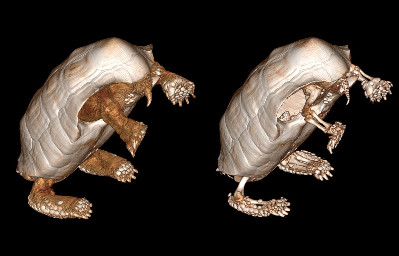
BENEATH THE SHELL
A CT scan of this tortoise shows its soft tissue (left) and bone structures (right), which helps veterinarians diagnose many different health conditions.
Tomographic reconstruction produces image “slices” (tomograms), which can be set for less than a millimeter—a staggering amount of detail is collected in seconds! Ben Nevitt, DVM, San Diego Zoo, described how he and his team had recently taken radiographs of a snake, which garnered six images from different angles, while a CT scan created 1,500 images of the reptile. “Getting this level of detail will really help us with the health of older animals,” he added. For instance, early arthritis is difficult to detect through radiographs, but the BodyTom reveals it effortlessly. Animals like turtles and tortoises also benefit, as CT scans can “see” through their shell to the bone and soft tissue beneath. This information helps veterinarians decide on the best course of action for the animal.

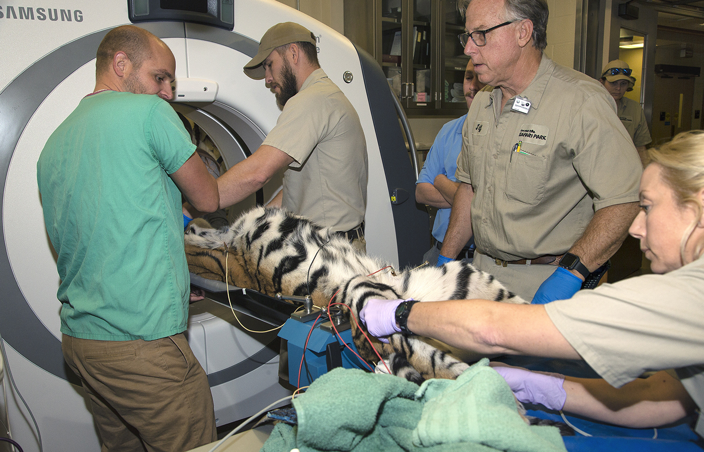
TENDER, THOROUGH TEAMWORK
This tree kangaroo is getting a full body CT scan to evaluate normal anatomy.
Elite Scanner
Approximately the dimension of a king-size mattress standing upright, the BodyTom Elite is a sleek, complex machine on “tank treads,” which ensures a smooth, consistent scan over the carbon fiber table holding the patient. It can be operated wirelessly, is fan-cooled, and can run on battery power alone. The scanner is easy to maneuver within the hospital—brackets flush to the floor mark a level, 8-by-8-foot square for the scanner to be positioned. There are five areas in the Paul Harter Veterinary Medical Center at the the Safari Park that can accommodate the CT scanner. Suites at the Zoo’s Jennings Veterinary Hospital are smaller, and the floors gently slope toward drains for cleaning purposes, but there is just enough level real estate in both surgical suites for this important diagnostic tool.
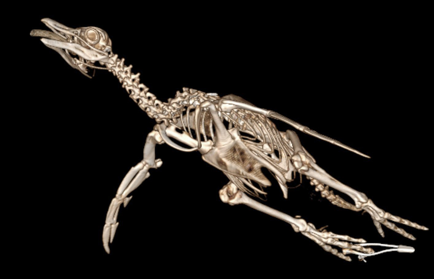
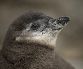
BIRD BONES
This African penguin’s CT scan reveals its skeleton in mind-boggling detail.
Just WOW!
CT scan images can be viewed in three formats: two-dimensional (2-D) view; multiplanar reconstruction (MPR) view, which generates sagittal (left to right), axial (front to back), and coronal (dorsal to ventral) slices; and three-dimensional (3-D) reconstructions of the patient. Dr. Kinney explained how important each different view is, because veterinarians “need to decide quickly on the best course of action, often while the animal is still under anesthesia.” He demonstrated how they can “digitally isolate certain anatomic structures to get better visualization of individual body regions,” which is fascinating and useful for diagnostics. Color can also be added to the images, to highlight individual anatomical structures.
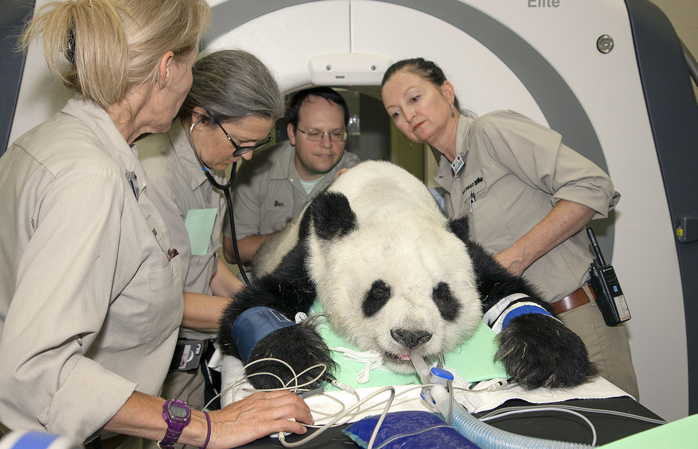
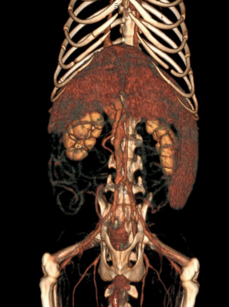
BEAR HEALTH
A giant panda is prepared for a diagnostic CT scan at the San Diego Zoo.
Two types of scan modes are used: axial and helical. For an axial scan, also called step-and-shoot scanning, the scan stops after each slice, and then moves on to the next slice. This takes more time, and the patient’s breathing can cause a “motion artifact” that distorts the slice. In contrast, helical scans (also called spiral scans) are continuous, rapid scans that collect thinner slices taken in a spiral fashion, but they can lose some detail. This mode is useful in catching clear pictures in between breaths or heartbeats in birds and other animals with rapid respiratory motion.
Veterinarians need all the information they can get to make the best treatment decision for the animal. For instance, an okapi with a joint lesion received a helical scan of its entire leg, then the team zoomed in with an axial scan of the joint to get a precise picture of its ailment. Images are placed in a “bone window” or a “soft tissue window” with the click of a mouse, and these different views are key to diagnosing disease and other health issues.
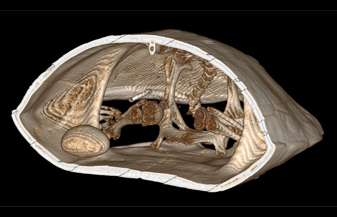
INSIDE SCOOP
This CT scan shows a developing egg (seen on the left) in the coelom of this turtle.
Eggs-act Science
One highlight for Dr. Kinney was earlier this spring, when a condor chick was not positioned correctly in the egg, which could have resulted in suffocation during the hatching process. Keepers brought the egg to the hospital, and a CT scan revealed exactly where its skull and beak were, indicating the best place for a helping pip hole. “An X-ray would have been less precise and required more mental gymnastics to get the right spot,” he said. Today, that condor chick is thriving with its parents at the Safari Park.
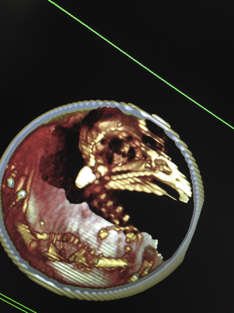
PEEK AND PIP
This California condor chick was not positioned correctly for hatching. A CT scan revealed the best location to help this chick start breaking through its shell.
Each patient study is uploaded to a computer server, so it is accessible between the Zoo and the Park, as well as off-site—the information can be shared with outside experts if needed. Sometimes a detailed evaluation is needed from a radiology specialist, who can see more subtleties in the image. “Our ability to read computed tomography images is improving with the more studies we view, but having experts available for consultation is a real asset,” Dr. Kinney explained. Another great feature is building a CT scan library of animals, which can create a baseline for a species and document changes in individuals over time. “We will have a metric for case follow-ups and repeat evaluations,” Dr. Kinney said. “This CT scanner helps us provide world-class medicine: improved health care, more accurate diagnoses, and superior treatment plans that ultimately result in better animal welfare,” he added. And that’s something we can all picture.