Zoo InternQuest is a seven-week career exploration program for San Diego County high school juniors and seniors. Students have the unique opportunity to meet professionals working for the San Diego Zoo, Safari Park, and Institute for Conservation Research, learn about jobs, and the blog about their experience online. Follow their adventures here on the Zoo’s website!
What happens when an animal is sick? What happens if they die? This week we met with pathology fellow Jenny Bernard and pathology resident Andrew Cartoceli at the Wildlife Disease Lab for San Diego Zoo Global. When an animal from the San Diego Zoo or San Diego Zoo’s Safari Park dies, they are brought to the Wildlife Disease Lab to be examined. Dr. Bernard and Dr. Cartoceti examine a variety of different animals, from small millipedes all the way up to the largest animals at the zoo; the elephants. However, they do not just examine deceased animals, they evaluate living ones that might have a disease that is affecting not only its own health, but the health of other animals around it.

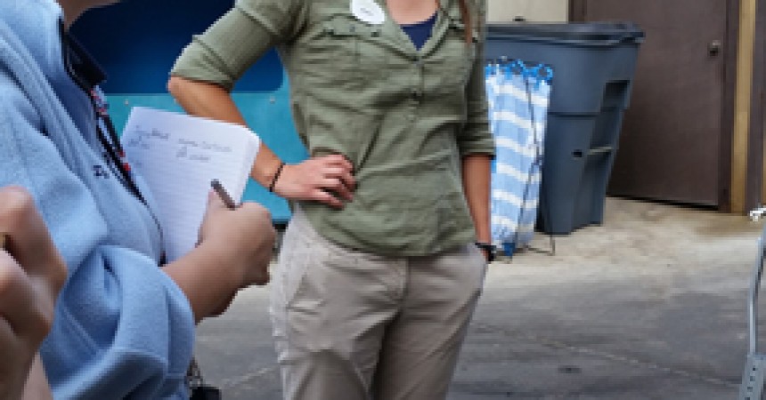
In the photograph above, Dr. Bernard tells us about the process taken when they receive a deceased animal. This process begins with a necropsy or gross examination. These terms are used when describing an animal autopsy. During the initial gross examination, they look at the anatomy of the entire animal. By doing this, they can see any abnormalities in the basic structure and functions of the internal organs. Next, the affected tissues are sent to the histology department. This is where the microscopic slides are prepared in order for Dr. Bernard and Dr. Cartoceti to look at the cells on a microscopic level. This is done to see if an infected area found in the gross necropsy was a virus, bacteria, or fungi.

Pathologist, Dr Bernard, analyzes tissues for disease and determines the results for a diagnosis. Contrary to popular belief, a pathologist is not like the television show CSI; the results are not always quick and conclusive. However, if tissue needs to be tested, the process can take several weeks and the results are not always clear.

After gross examinations some of the organs, body parts, and tissues are preserved for education purposes. The body parts and tissues not needed for education purposes are put through a machine known as the tissue digester. The digester breaks down tissues into liquids and bones into a chalky-like substance as seen above. The chalky bones can then be used as fertilizer throughout the San Diego Zoo.

The machine pictured above is used to stain the tissues on the microscopic slides to show the different parts of the cell. The stain used depends on if they are testing for bacteria, fungi, or a virus in the animal’s cells. Histologists can also use a special stain that will show a specific part of the cell they might be interested in looking at.

Here are blue-stained slides, one of the “typical” colors used to differentiate between organelles. After they are stained, the slides can be looked at under a microscope to see various abnormalities in question. If it is bacteria or fungus, Dr. Bernard and Dr. Cartoceti would be able to identify it because of the shape of the organism. However, sometimes pathologists are unable to identify a specific pathogen, but they can eliminate contagious diseases which could potentially affect the other animals in the collection.

After the slides are made in histology, they are sent to Dr. Cartoceti and Dr. Bernard to be analyzed by looking at them under the microscope. Through these slides, they can identify pathogens that were inside the animal. Dr. Cartoceti was able to show us some of the cases he worked on. He pulled up a slide under a microscope of an actual bacteria and fungi and showed us how to find what the infection was and how it changed the anatomy when it was alive.
Isabella, Photography Team
Fall Session 2014